
In a recent article, I discussed the often neglected concept of constitutional archetypes. Briefly, this concept encapsulates the idea that while everyone is different, there are common patterns in those differences, which if recognized can be used to tailor a treatment to what best serves the archetype that patient belongs to.
For example, the elderly respond very differently to pharmaceutical drugs than the young (which leads to them frequently being severely injured — best show by a study that found simply reducing the number of pharmaceutical drugs they were taking by a third reduced their risk of dying by 23%).
Unfortunately, while there are many common demographics (e.g., women) that have predictable reactions to pharmaceuticals, in most cases, the differences in how they respond to drugs are not considered. Instead everyone typically gets the same standardized dosage, frequently leading to disastrous consequences that could have been predicted had their archetype been considered from the start.
Note: The tragic (and frequently preventable) consequences that follow SSRI usage are one such example (discussed further here).
One of the most important unrecognized demographics are the “sensitive patients,” individuals who are typically much more intuitively sensitive to the subtle qualities in their environment (e.g., they viscerally perceive the unsaid emotions of others), but simultaneously are also much more sensitive to being injured by pharmaceuticals.
In turn, I’ve lost count of how many of these people I’ve met (e.g., many are close friends) and the way that they are frequently treated by the medical system deeply bothers me.
Note: This constitutional archetype is discussed further here.
Ligamentous Laxity
At this point, I often immediately recognize the sensitive patients by their spiritual presence, their psychological demeanor and their body shape. One of the frequent physical traits I associate with this archetype is a distinct pattern of movement (which is often graceful) that I believe results from laxity in their ligaments.
Since the ligaments stabilize the joints of the body by constraining their maximum range of motion, once the ligaments weaken, their laxity (looseness) frequently causes the maximum range of motion of the joint to increase.

Individuals with ligamentous laxity have certain advantages. For example, they are normally much more flexible (e.g., they can stretch their body much further), and often become dancers because of the mobility afforded to their body.
Conversely, ligamentous laxity also creates a variety of issues for those patients. The most common one is that since their body uses the ligaments to stabilize itself when a force enters it, once the ligaments weaken, things will frequently move inside the individual more than they should in response to those forces.
For example, individuals with ligamentous laxity tend to be more susceptible to developing chronic injuries from car accidents or other physical traumas. Likewise, their joints “pop” easily, but if they get forceful adjustments to reset the position of their bones, the bones will often go back out of place (since the stability which would otherwise maintain that structure is lacking).
Note: I have seen numerous patients with ligamentous laxity that had far too many forceful adjustments that eventually created issues for them. This normally occurs either because the repeated adjustments (performed due to the patient’s pain rapidly returning after each adjustment) further weakened their ligaments or because the joint exceeded its expected range of motion during a thrust and the bone ended up in a position which created other issues for the body.
Similarly, individuals with ligamentous laxity (especially when it is significant) tend to develop a variety of issues that are ultimately treated with surgery. Unfortunately, due to being more “sensitive” and having an impaired ability to heal following the surgery, they frequently suffer significant complications from those surgeries (e.g., there are many stories of this in the Ehlers-Danlos syndromes [EDS] support groups).
Note: Since ligaments are composed of collagen, impaired collagen production often gives rise to ligamentous laxity. Likewise, since collagen production is needed to heal from surgeries, individuals with impaired collagen production have greater difficulty recovering from surgery.
Ligamentous laxity can also create a variety of other issues throughout the body (e.g., all sorts of gastorintestinal problems). One of the least appreciated consequences of hypermobility is that the body depends upon ligamentous tension for proprioceptive feedback (knowing where the body is in space), and as a result, once the ligaments become lax, hypermobile patients lose some of the general awareness that allows us to subconsciously navigate through the world.
For example, EDS is known to be associated with impaired balance and an increased likelihood of falling.
However, it’s much less appreciated that this lack of proprioceptive feedback can give rise to a general sense of anxiety because the ground does not feel as stable below one’s feet — something which is difficult to appreciate unless one has experienced it directly (e.g., I’ve met people who easily survived a major earthquake but were psychologically disturbed for years after as a result of them briefly losing their sense of the ground which they had always previously taken for granted).
What Causes Ligamentous Laxity?
A variety of processes can cause ligamentous laxity. Frequently, a combination of these are at work (e.g., someone with a pre-existing weakness in a ligament is more likely to have something injurious to a ligament create a chronically lax ligament).
• Physical injuries — Ligamentous weakness is frequently caused by a chronic injury to a ligament (or an injury which did not properly heal). For example, when the ankle is injured from a sprain, the ligament that keeps the ankle from turning inwards gets damaged, predisposing us to an unstable ankle and future ankle injuries until the ligament is repaired and strengthened.

• Pharmaceutical injuries — In addition to physical injuries, biochemical injuries can also occur. This is best known to occur following the use of anti-inflammatory medications. For example, NSAIDS like Ibuprofen (which are typically the go to for “treating” sports injuries) in addition to reducing the discomfort of an ankle injury also weaken the ligaments (as by suppressing the inflammatory process it also suppresses the healing process).
This results in sprains that are treated with NSAIDs often being predisposed to future injuries (since the stability given to the joint by those ligaments is partially lost until something like a regenerative therapy is given to repair that ligament) — which in the case of ankle sprains is something I and colleagues frequently see in our patients.
One of the most common therapies given to treat joint pain are injected steroids, which, being powerful anti-inflammatories, are often initially effective in making the patient feel better. Unfortunately, injected steroids are also weaken ligaments at the site where they are injected, particularly when they are given multiple times.
Since joint inflammation (and pain) often is a result of improper joint stabilization by a ligament (leading to the joint being worn down by inappropriate pressures), this treatment approach results in patients needing more and more injections as their ligaments further weaken.
Note: This is also a common issue for those with chronic neck or back pain and unfortunately often results in them often “needing” to get a surgery to stabilize the spine — which often makes things even worse (that subject and alternative approaches to treating spinal pain are discussed here).
Additionally certain more toxic pharmaceuticals can also weaken the ligaments. In addition to steroids, I have most commonly observed this after the someone suffers an injury from a more toxic pharmaceutical (e.g., this is well known to occur from fluoroquinolone antibiotic like ciprofloxacin but I’ve also seen it after an accutane injury).
Presently, the best explanation I’ve come up for this is that the body is continually rebuilding and remodeling its structural tissues to meet the needs of the environment. For example, the bones depend on the weight of gravity to signal growth, so when astronauts are in space for prolonged periods of time, their bones significantly weaken (and may break once they return to Earth’s gravity) unless the astronauts also do special exercises in space.
In addition to bones, the collagen of the body also continually rebuilds and remodels itself. Since collagen production is an energy intensive process, if the mitochondria get injured (which fluoroquinolone antibiotics are notorious for causing), over time the connective tissue in the body will weaken until it fails due to it longer being able to accommodate a force input (e.g., sudden tendon ruptures are very common after fluoroquinolone usage).
• Constitutional predispositions — EDS and Marfan Syndrome are the two classic medical conditions that are associated with ligamentous laxity. Both of these conditions are considered to be rare diseases, with EDS being estimated to affect between 1 in 5000 to 1 in 100,000 patients (depending on its severity and the source of the estimate), while Marfan Syndrome is estimated to affect 1 in 5000 patients.
Note: One recent paper found that the prevalence of diagnosed EDS was 1 in 500 and many believe it is even more common (e.g., I’ve met dozens of people who have appear to have EDS or an EDS-like syndrome).
In addition to EDS, there is also a self-explanatory condition known as “generalized joint hypermobility,” (GJH) which is found to affect around 12.5% of the population and predisposes them to musculoskeletal pain and injury. At this point I suspect that both this condition and EDS are under-diagnosed and that a spectrum exists between overt EDS and GJH.
• Functional predispositions — It some cases you can watch a ligamentous laxity rapidly onset within the body. The most classic example results from the hormone relaxin (released during pregnancy), which weakens the ligaments of the body so that the pelvis becomes able to stretch and accommodate the birthing process.
Because of this increased hyper-mobility, pregnant women become more predisposed to physical injuries (and developing musculoskeletal pain from the weight their baby).
Note: Other hormones the female body secretes counteract the effects of relaxin, especially once the pregnancy is complete, and in turn, those with pre-existing hormonal imbalances are significantly more susceptible to musculoskeletal issues both during and after a pregnancy.
Likewise, having pre-existing hormonal imbalances (due to the ligamentous weakness they create) can make women much more susceptible to musculoskeletal issues (e.g., in the feet), particularly after menopause.
In my eyes the most important functional predisposition of ligamentous integrity is nutritional status. While a variety of things can affect ligamentous integrity, we’ve found the often forgotten metal manganese (Mn) plays one of the most important roles. In turn, it is extremely common to find people who:
◦ Live in areas with lower manganese levels in the soil also appear to have higher rates of ligamentous laxity.
◦Have ligamentous laxity that gradually improves when they appropriately take manganese for a prolonged period.
◦ Have a predisposition to ligamentous laxity (e.g., EDS or a hypermobility syndrome) be tipped over the edge by a manganese deficiency, and likewise have patients with existing hypermobility significantly improve from manganese supplementation.
Note: Our preferred approaches for supplementing manganese are discussed in more detail here.
Since manganese deficiency is such a common issue now, we’ve put a lot of thought into why we see it so frequently. Presently, we have three leading explanations:
◦ First, the topsoil has been heavily depleted of essential minerals by our modern farming practices. This creates a variety of issues (and likewise significant improvements are seen in the plants grown in remineralized soil and in the humans who consume those plants).
◦ Glyphosate (Roundup), a toxic but widely used herbicide, has a high affinity for chelating (trapping) certain minerals, once of which is manganese. Because of this, I believe the continually increasing levels of glyphosate in the environment have contributed to the current widespread deficiency of manganese.
◦ Bacteria require iron for their metabolism (and in turn one of the defensive mechanisms the body has to stop an infection is to sequester iron so bacteria cannot reproduce). The bacteria which causes lyme disease has an unusual adaptation, which to my knowledge is unique in nature (which raises interesting questions about where Lyme came from).
It uses manganese instead of iron, which thereby protects it from that defense mechanism, and we have long suspected it also causes individuals infected with the bacteria (including those with “silent” infections) to become manganese deficient.
Note: Manganese is often considered to be a toxic element (as it is associated with the neurological issues that follow chronic exposure to welding gas). I always thought this was strange as manganese is a necessary cofactor for numerous critical enzymes in the body (e.g., some of those within the mitochondria).
Presently, I believe this is because the toxic form of manganese (e.g., that found in welding gas) is Mn3+, whereas the form that helps people found in the dietary supplements (and that typically tests well for patients) is Mn2+.
In turn, I suspect that for some reason, Mn3+ impairs the function of essential enzymes that depend upon Mn2+. Alternatively, a hypothesis has been put forward (with data to support it) that manganese, while essential, also becomes toxic at high doses.
Ligamentous Laxity and Vaccine Injuries
Prior to COVID, the HPV vaccine Gardasil was considered by many to be the most dangerous vaccine released upon the general population (a strong case could also be made that it was either the original DPT vaccine or the original smallpox vaccine, however by the time Gardasil emerged, those atrocious vaccines had mostly faded into memory).
Gardasil caused a variety of debilitating conditions (e.g., constant fatigue, cognitive impairment, a variety of autoimmune conditions and widespread neurologic dysregulation — things also frequently seen after the COVID-19 vaccination).
At some point, holistic physicians began noticing patients (particularly women) with existing ligamentous laxity were much more likely to develop Gardasil injuries (and in turn a few researchers proposed genetic variations to account for this). At the time, I assumed this correlation was due to:
• Individuals with ligamentous laxity also being more likely to be the more sensitive patients.
• Ligamentous laxity having a longstanding association with mast cell disorders, a condition which makes one hypersensitive to a variety of substances (e.g., drugs or vaccines).
Note: Mast cell disorders are frequently observed to emerge in patients who develop COVID-19 vaccine injuries.
However, a few months ago I learned of a novel hypothesis put forward by one of the leading doctors treating COVID-19 vaccine injuries.
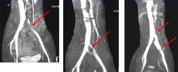
Iliac Vein Compression
Dr. Jordan Vaughan’s key points from this twelve minute video (and my comments on them) are as follows:
• Many of the debilitating complications of the COVID-19 vaccine appear to result from the iliac vein (which governs venous return from the legs and pelvis) becoming compressed and its blood supply becoming significantly obstructed (which in turn gives rise to numerous characteristic symptoms that can be recognized once one knows to look for them).
More importantly, this compression can often onset quite suddenly and patients remember the exact moment that compression occurred.
• The blood flow obstruction created by an iliac vein compression appears to cause a variety of conditions which could be analogized to “putrid blood,” such as blood clots throughout the body and mast cell activation syndrome.
Note: Over a century ago, quite a few of the early pioneers in the medical field also noted that compression of the iliac region predisposed patients to a variety of illnesses they commonly attributed to the blood or lymph in the pelvis “fermenting.”
• A condition known as May-Thurner syndrome affects slightly over 20% of the population. In it, the (high pressure) right common iliac artery overlies and compresses the (low pressure) left common iliac vein against the lumbar spine.

Normally, individuals with this syndrome do not notice it, but it some cases it can predispose them to unexpected blood clots (due to venous return being obstructed).
• Dr. Vaughan believes that the (well-documented) damage the spike protein causes to walls of the blood vessels weakens the iliac vein enough that pressure from the iliac artery can cause it to collapse. In turn, he has collected compelling radiography to prove it.
• If an iliac vein compression is treated with anticoagulation, this typically improves the symptoms of it, but once the anticoagulants are stopped, the symptoms return (as the source of the clotting has not been addressed). For this reason, iliac vein compressions are often treated with stenting, which Dr. Vaughan reports frequently significantly benefits patients.
Note: Years ago, a (still) famous integrative physician found that he could dramatically improve a variety of complex neuroimmune conditions (e.g., Lyme disease, fibromyalgia and chronic fatigue syndrome) by stenting the jugular vein, but eventually had to stop because the government stopped the practice.
After I learned about this, I asked about and discovered visceral therapists who do manual work on the arteries and veins (which allowed them to decompress the jugular vein without a stent) had similar results to that physician.
Once I heard Dr. Vaughan’s lecture, I realized this hypothesis likewise explained many of the successes they had shared in the treatment of vaccine injured patients as the manual therapists I spoke to, either intentionally or unintentionally had partially (or fully) decompressed the iliac vein without the use of a stent.
As stenting (like any other surgical procedure) carries some degree of risk, my first line therapy for vaccine injured patients with signs of an iliac vein compression is thus the hands on approach.
After seeing Dr. Vaughan’s lecture, I realized it tied together a lot of observations I had made over the years.
Zeta Potential
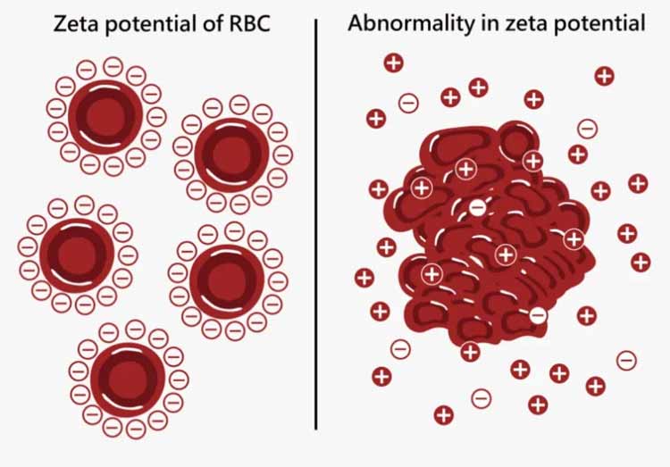
Zeta potential (described in more detail here) quantifies the strength of the net electrical charge between particles colloidally suspended in a solution. Since almost all liquid systems in nature are colloidal suspensions, zeta potential is hugely consequential since it determines if the components of the system will remain apart (dispersed) or clump together (agglomerate), at which point they stop being suspended in the surrounding solution.
Since a variety of things encourage agglomeration (e.g., gravitational separation and the van der Waals force), for a colloidal system to remain stable, the existing zeta potential must be sufficient to overcome those forces.
This occurs by having the suspended particles share the same electrical charge (as opposites repel), which in almost all cases is negative (which I believe is due to water frequently forming a phase [commonly described as a liquid crystalline phase] that creates a layer which coats suspended particles with a negatively charged lattice of H3O2– ions).
Decades ago, Andrew Moulden (building upon the work of Thomas Riddick) made the extraordinary discovery that many of the frequently observed harms of vaccination could be traced to them flooding the body with positive charges that caused its blood cells to clump together, forming microclots that were too small to see with conventional imaging (e.g., a MRI — although these clots can be seen with specialized microscopes) but large enough to cause microstrokes in critical smaller arteries throughout the brain.
Note: Aluminum (which is routinely added to vaccines), due to its high positive charge density, is one of the most powerful agents that exists for destroying colloidal stability (e.g., most sewage is treated [flocculated] with aluminum to separate organic matter present from water).
In turn, Moulden was able to demonstrate that the cranial nerves (IV, VI and VII) that were particularly sensitive to microstrokes (e.g., the one that let’s your eyes look to the sides or the one that controls facial muscle tone) would frequently be impaired in vaccine injuries, and that their impairment frequently served as a sign there were other significant neurological issues resulting from microclotting within other parts of the brain (e.g., he correlated it to many cases of autism).
Note: A lot of people I know developed horizontal eye tracking issues (a CN VI palsy) after being vaccinated for COVID.
While I frequently consider zeta potential both in vaccine injuries and many infectious diseases (e.g., septic patients and patients who are severely ill with the flu often have significantly impaired zeta potential), my emphasis on it became much greater during COVID as from the start I noticed it was doing some very unusual things to patients I associated with significant impairment of the physiologic zeta potential.
Note: In addition to impaired zeta potential causing microclots, it can also cause agglomeration in the other fluids of the body (e.g., the lymphatics), something we saw throughout COVID-19.
Suspecting something in the virus (which was not in the original SARS virus) had to carry a very concentrated positive charge, I started looking through each protein in the virus. Before long, I realized the spike protein was the most likely candidate (as it was on the outside of the virus and had a cluster of positive charges not found in the original SARS virus).
In turn, I became quite concerned when I learned that the COVID vaccine would mass produce the spike protein throughout the body, and since that time, I’ve noticed many of the vaccine’s side effects mirror what would be expected during a significant impairment of the physiologic zeta potential.
Note: Not too long ago, a paper was published that demonstrates the spike protein does indeed impair the physiologic zeta potential.
Zeta Potential and Venous Compression
Dr. Vaughan made me realize there was a critical correlate of zeta potential I had not considered before.
In addition to the mutual negative charges causing those particles to be pushed away from each other, they also cause the walls of their container to pushed apart because:
- The blood vessels are also lined with a negatively charged lattice of H3O2– ions.
- The particles within the vessel, due to their mutual repulsion, cannot be compressed (which in turn prevents the walls of the vessel from compressing inward).
Over the years, I have observed that many of the challenging chronic neuroimmune illnesses that are associated with both poor circulation and mast cell activation syndrome (e.g., lyme disease and mold toxicity) also have unique characteristics that impair zeta potential. In turn, I find that attempts to treat these diseases fail unless something is also done to mitigate their adverse effects on zeta potential (especially within the lymphatic system).
Note: These are the same conditions that were previously found to respond to stenting of the jugular vein (the vein in the neck that drains blood and waste products from it).
Likewise, Gardasil (which was associated with a variety of circulatory impairments) has also been observed to trigger mast cell activation syndrome. In turn, many of the issues with Gardasil came from the fact the human body did not want to develop a strong response to the HPV vaccine’s antigen (which I suspect was due to it being too similar to human tissue), something which was not acceptable for Merck.
This “problem” was solved by using a very powerful aluminum adjuvant — which got Gardasil approved, but as shown in the trials (where the adjuvant alone was used as a “placebo”) also caused a wide range of severe issues for the vaccine recipients. In turn, when Moulden (and those who followed him) looked at the HPV vaccine, they observed that it was remarkably effective at disrupting the physiologic zeta potential.
Note: I believe the vaccines which most effectively disrupted the physiologic zeta potential were:
• The original smallpox vaccine.
• The anthrax vaccine (which was only given to the military but Moulden nonetheless focused on due to how overt many of its effects on zeta potential were).
• The now banned DTwP (we use the DPaT now and ship DTwP to the third world).
• The COVID-19 vaccine.
In short, I now believe a key reason why I often see so much improvement in venous circulation after treatments are provided to restore the zeta potential of the body is because doing so “re-expands” the compressible vessels of the body. I suspect this also holds true for the lymphatic vessels. Additionally, we frequently find that treating the zeta potential of the body is critical for healing COVID vaccine injuries.
Note: Since microcirculation largely depends on liquid crystalline water (H3O2-) driving the flow of the fluids within a vessel (explained further here), once the vessels becomes compressed together, in addition to vessel becoming blocked, the vessel’s contribution to the inherent movement of fluid through the vessels is lost as well.
Conclusion
One of the most challenging things in medicine is how interconnected the systems within it are, to the point it is often very difficult to describe a single part of it in isolation.
In this article, I attempted to show how (the sadly common) conditions which predispose one to ligamentous laxity also predispose one to mast cell disorders, a weakened lining of the blood vessels, and venous collapse (and likely lymphatic vessel collapse — as the lymph is also a low pressure system).
Note: The hypermobility syndromes are also associated with circulatory issues. Marfan Syndrome for example is associated with a weakening (and eventual bulge or tear) of the aorta (which is very bad) along with circulatory impairments throughout the body.
In many cases, we can compensate for that structural impairment of the vessels (e.g., because of the inherent force that drives fluid through the vessels and because of the mutually repulsive forces that keep the blood vessels open). However, once zeta potential is partially lost (which impairs circulatory movement and allows the vessels to collapse), the underlying impairment within the vessels can reach a tipping point where serious issues follow.
This in turn I would argue is a key reason why certain individuals are so much more susceptible to injuries which impair the physiologic zeta potential.
Note: Patients who already have an impaired zeta potential are also more sensitive to successive worsening of their zeta potential. This for example is why the elderly (who have often lost part of their zeta potential due the kidney’s ability to maintain it declining with age) are often so much more susceptible to the effects of an infection (e.g., the flu or a common infection). Likewise, it also helps to explain why patients often worsen with each successive COVID vaccination.
I sincerely thank you for taking the time to read this article and consider the ideas put forward in it as I believe they are critical to unravelling the challenging puzzle before us now.
A Note From Dr. Mercola About the Author
A Midwestern Doctor (AMD) is a board-certified physician in the Midwest and a longtime reader of Mercola.com. I appreciate his exceptional insight on a wide range of topics and I’m grateful to share them. I also respect his desire to remain anonymous as he is still on the front lines treating patients. To find more of AMD’s work, be sure to check out The Forgotten Side of Medicine on Substack.